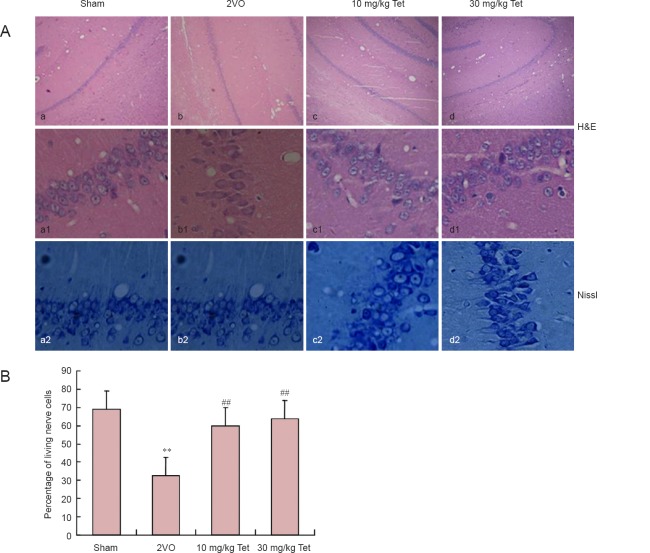
Figure 2.
Effect of Tet on hippocampal CA1 pathology in rat models of vascular dementia.
(A) H&E (upper panel, 40× magnification; middle panel, 200× magnification) and Nissl staining (lower panel, 200× magnification) of hippocampal CA1 showing atrophic or dying cells (arrows). Scale bars: Upper panel, 5 μm; middle and lower panels, 50 μm. (B) Percentage of living cells in CA1 (mean ± SD, n = 3 rats per group). **P < 0.01, vs. sham group; ##P < 0.01, vs. 2VO group (one-way analysis of variance and Bonferroni post hoc test). Tet: Tetrandrine; 2VO: two-vessel occlusion; H&E: hematoxylin-eosin.

