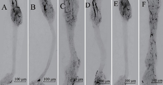
Figure 1.

Vascularized images of acellular nerves.
In the control group, acellular nerve grafts alone were transplanted. In the test group, acellular nerve grafts were transplanted and daily intraperitoneal injections of cartilage oligomeric matrix protein-angiopoietin-1 were performed. Vessels grew into the acellular nerve graft from both anastomotic ends beginning at day 7 (A, D) and gradually covering the entire graft at day 21 (C, F) in both the control and test groups. The velocity of vascularization in the test group (D–F) was faster than that in the control group (A–C). Vessels are indicated by the black areas. Scale bars: 100 μm.
