FIG. 3.
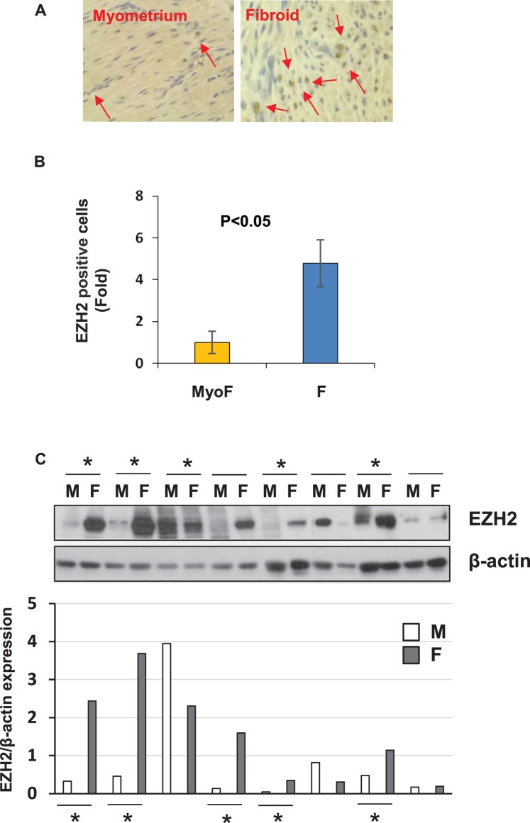
Expression of EZH2 expression in fibroid tissues and adjacent myometrium. A) Immunohistochemistry of EZH2 expression in fibroid and adjacent tissues. Arrows indicate EZH2-positive cells (magnification ×400). B) Semiquantitative analysis of EZH2-positive cells in myometrium and fibroids from fibroid and matched myometrium samples (n = 2). C) Western blot analysis of EZH2 in fibroid and adjacent tissues from African American women (n = 8). Total lysates from fibroids and adjacent myometrial tissues were extracted and subjected to Western blot analysis using antibody against EZH2. βactin was used as an endogenous control. Asterisks indicate the upregulation of EZH2 in fibroid tissues as compared to adjacent myometrium tissues. EZH2 protein bands were quantified and normalized to βactin, and relative values were used to generate data graphs (C, bottom panel). F, uterine fibroid; M, adjacent myometrial tissue.

