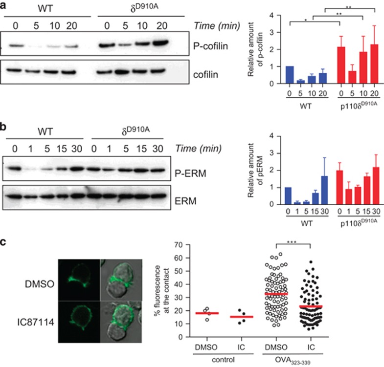
Figure 2.
PI3Kδ controls cofilin and ERM regulation and cell polarisation. WT and p110δD910A CD4+ T cells were activated with anti-CD3 and anti-CD28 mAbs for the indicated times and lysed. Lysates were then analysed with the indicated antibodies in order to measure cofilin activity (a) or ERM phosphorylation (b). For quantification, relative amounts of phospho-cofilin or phospho-ERM were normalised to the total amount of cofilin or ERM. P-values were calculated using a two-way repeated-measures ANOVA. *P<0.05. Data presented are representative of three independent experiments. (c) Actin-GFP expressing OT2 CD4+ T cells were pre-treated with DMSO or the p110δ specific inhibitor IC87114 before being incubated with B cells loaded with OVA323-339 peptide. After 30 min, cells were fixed and the intensity of the fluorescence of actin-GFP at the contact zone measured. Data are representative of at least three independent experiments.

