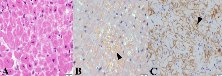
Figure 5.
Histological findings in myocardium from the right ventricle. An infiltration of lymphocytes was observed in the biopsied specimen. There were focal atrophic myocardial changes accompanied by deposition of eosinophilic materials. (A) Deposition of eosinophilic materials can be observed in the myocardial interstitium (hematoxylin and eosin, objective lens ×60). (B) A bright greenish yellow color (arrowhead) after staining with Congo red can be seen under polarized light (Congo red, objective lens ×60). (C) A strongly positive immunohistological reaction for immunoglobulin λ-chain can be seen in the myocardial interstitium (arrowhead, dark-brown color) (immunoperoxidase reaction for λ-chain, objective lens ×60).

