Figure 2.
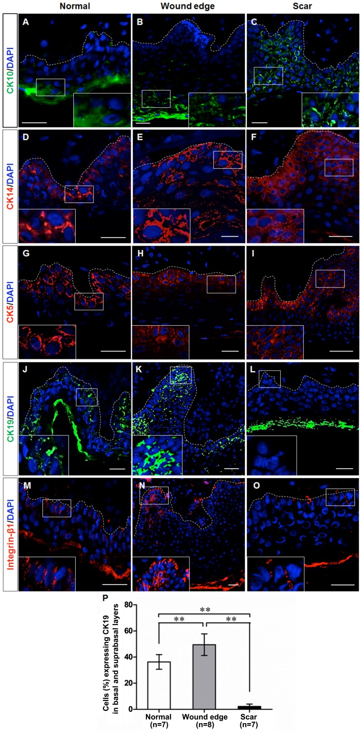
Cell marker expression in epidermal keratinocytes in normal, wound edge and hypertrophic scar tissue. Representative fluorescence images showing the localization of CK10, CK14, CK5, CK19 and integrin-β1 in the sections of normal, wound edge and scar tissues. (A–C) CK10, a marker of differentiated keratinocytes, was expressed in the outer layer of the epidermis of (A) normal and (B) wound edge tissues, and in (C) the suprabasal, terminally differentiating cells in the epidermis of hypertrophic scar tissues. (D–F) CK14 was expressed in both basal and suprabasal layers in the stratified epidermis of all three tissues. In particular, extensive distribution of CK14 was detectable in (F) the multilayered epidermis of the scar tissue. (G–I) The distribution patterns of CK5 were similar to those of CK14, which indicated an increased number of proliferating cells in the hyperproliferative epidermis. (J–O) CK19 and integrin-β1 were expressed in the basal layer of (J and M) normal epidermis, and in the basal and suprabasal layers of (K and N) wound edge epidermis, but were not detected in (L and O) the epidermis of hypertrophic scar tissues. White dotted lines denote the basement membrane, which separated the epidermis and dermis. Insets show a close-up view of area within the white markings. All scale bars represent 25 μm. (P) Statistical analysis of CK19-expressing cells in the basal and suprabasal layers in normal, wound edge and hypertrophic scar tissues. Data are the means ± SD. **P<0.01.

