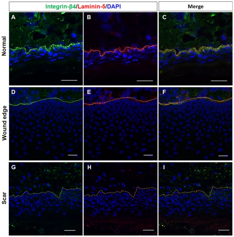
Figure 4.
Representative fluorescence images showing the double-labelling of laminin-5 and integrin-β4 in normal, wound edge and hypertrophic scar tissue. (A–I) The distribution of laminin-5 and integrin-β4 was detected in the basement membrane (BM) area in both (A–C) normal and (D–F) wound edge skin tissues. (G–I) The formation of BM-like structures in the absence of laminin-5 and integrin-β4 expression was observed in the epidermis of hypertrophic scar tissues. White dotted lines denote the basement membrane, which separated the epidermis and dermis. All scale bars represent 25 μm.

