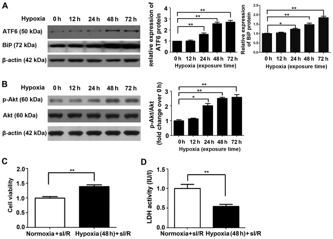
Figure 4.
Cleaved activating transcription factor 6 (ATF6) and Akt expression and effects on simulated ischemia/reperfusion (sI/R) injury induced in H9c2 cells subjected to mild hypoxia. (A) Representative expression of cleaved ATF6 and β-actin protein in H9c2 cells cultured under normoxic or hypoxic conditions for 0, 12, 24, 48 and 72 h (left); quantification of cleaved ATF6 and binding immunoglobulin protein (BiP) protein levels (normalized to β-actin protein levels) (right, *P<0.05 vs. 0 h; **P<0.01 vs. 0 h). (B) Representative expression of Akt, p-Akt and β-actin protein in H9c2 cells cultured under normoxic or hypoxic conditions for 0, 12, 24, 48 and 72 h (left); quantification of p-Akt protein levels (normalized to total Akt protein levels) (right, *P<0.05 vs. 0 h; **P<0.01 vs. 0 h). (C) Effect of mild hypoxia on the viability of H9c2 cells subjected to ischemia (1 h) followed by reperfusion (3 h). Representative data from five independent experiments are presented as the means ± SEM (**P<0.01 vs. normoxia group). (D) Effect of chronic mild hypoxia on lactate dehydrogenase (LDH) activity in H9c2 cells subjected to ischemia (1 h) followed by reperfusion (3 h). Representative data from five independent experiments are presented as the means ± SEM (**P<0.01 vs. normoxia group).

