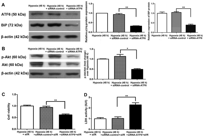
Figure 5.
Cleaved activating transcription factor 6 (ATF6) and Akt expression and effects on simulated ischemia/reperfusion (sI/R) injury induced in siRNA-ATF6-transfected H9c2 cells subjected to mild hypoxia. (A) Representative expression of cleaved ATF6 and β-actin in siRNA-ATF6-transfected H9c2 cells cultured under hypoxic conditions for 48 h (left); quantification of cleaved ATF6 and binding immunoglobulin protein (BiP) protein levels (normalized to β-actin protein levels) (right, **P<0.01 vs. siRNA-control). (B) Representative expression of Akt, p-Akt and β-actin proteins in siRNA-ATF6-transfected H9c2 cells cultured under hypoxic conditions for 48 h (left); quantification of p-Akt protein levels (normalized to total Akt protein levels) (right, *P<0.05 vs. siRNA-control). (C) Effect of mild hypoxia on the viability of siRNA-ATF6-transfected H9c2 cells subjected to ischemia (1 h) followed by reperfusion (3 h). Representative data from five independent experiments are presented as the means ± SEM (**P<0.01 vs. siRNA-control). (D) Effect of chronic mild hypoxia on lactate dehydrogenase (LDH) activity of siRNA-ATF6-transfected H9c2 cells subjected to ischemia (1 h) followed by reperfusion (3 h). Representative data from five independent experiments are presented as the means ± SEM (**P<0.01 vs. siRNA-control).

