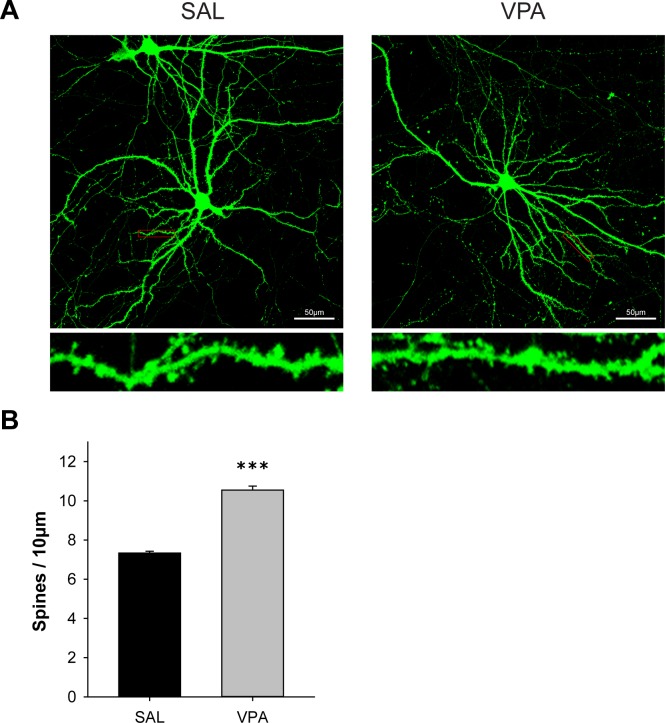
Fig 3. VPA mice showed increased dendritic spine density.
Cortical neuron cultures were obtained on E18 from fetuses prenatally exposed to SAL or VPA on E13, and the cells were maintained until DIV 19–20. (n = 3 mice for each group, n = 3–6 cells for each mouse, 3–6 branches were analyzed per cell.) (A) Representative fluorescence images of dendritic spines in primary neuron cultures. (B) Quantification of spine density of spine density on secondary dendrites 50–100 μm away from the center of the soma. VPA mice exhibited a significant increase in spine density. (* significantly different among groups, *** p < 0.001, n = 18 for SAL, n = 13 for VPA).

