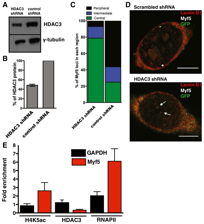
Fig. 4.
HDAC3 knockdown reduces localization of Myf5 with the nuclear lamina. a Wildtype myogenic progenitors treated with control or HDAC3 shRNA were separated by SDS-PAGE and western blotted with antibodies against HDAC3 and γ-tubulin. b Quantification of HDAC3 levels in HDAC-downregulated progenitors was normalized to γ-tubulin and control shRNA-treated cells. Error bars are S.E.M. n=3. c Quantification of Myf5 localization in control and HDAC3-downregulated myogenic progenitors. n=50. p<0.05. Peripheral is ≤1 μm from the nuclear lamina. Intermediate is between 1 μm and 2 μm from the nuclear lamina. Central is ≥2 μm from the nuclear lamina. d 3D-ImmunoFISH of Myf5 localization in control and HDAC3-downregulated myogenic progenitors co-transfected with GFP. GFP is green, Lamin B1 is red, Myf5 is white. Arrows indicate Myf5 loci. Scale bars are 5 μm. e ChIP-qPCR of Myf5 and GAPDH promoters with antibodies against RNA Polymerase II (RNAPII), H4K5ac, and HDAC3 in wildtype and emerin-null proliferating myogenic progenitors. Fold-enrichment was determined by comparing emerin-null cells to wildtype. p≤0.05 between wildtype and emerin-null for all three antibodies. Error bars represent S.E.M. n=3

