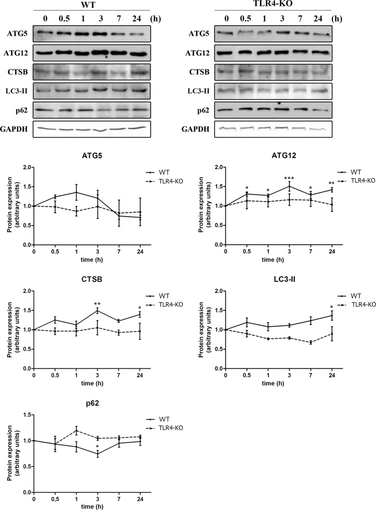
Fig 1. TLR4 participates in the ethanol-induced overexpression of several autophagic proteins in cortical astroglial cells.
Immunoblot analysis and quantification of ATG5, ATG12, cathepsin B, LC3-II and p62 in cell extracts of ethanol (50 mM)-treated cells at different time points (0, 0.5, 1, 3, 7 and 24 h). Values represent mean ± SEM, n = 12–15 independent experiments. * p < 0.05, ** p < 0.01, *** p < 0.001 compared with the untreated WT or TLR4-KO value. Blots were stripped, and the total quantity of GAPDH was also assessed. A representative immunoblot of each protein is shown.

