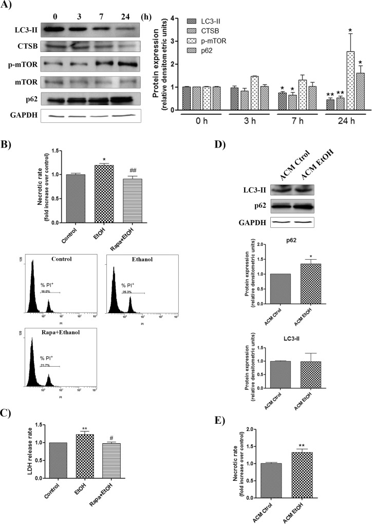
Fig 7. Ethanol down-regulates the autophagy pathway in cortical neurons and also causes necrotic cell death.
(A) Immunoblot analysis and quantification of LC3-II, p62, CTSB and p-mTOR in the cell extracts of the ethanol (50 mM)-treated WT neurons at different time points (0, 3, 7 and 24 h). Values represent mean ± SEM, n = 8–10 independent experiments. * p < 0.05, ** p < 0.01 compared with the untreated WT value. Blots were stripped, and the total quantity of GAPDH and mTOR was also assessed. A representative immunoblot of each protein is shown. (B) Autophagy enhancer rapamycin was incubated in the WT neurons for 1 h before initiating the 24 h ethanol treatment. The necrotic cell death analysis of cortical neurons was analyzed by flow cytometry. Data represent mean ± SEM, n = 8–10 independent experiments. * p < 0.05 as compared to the control group, ## p < 0.01 as compared to the ethanol-treated group. (C) LDH activity was measured in the supernatant of cortical neurons, untreated or treated with ethanol and rapamycin (100 nM) plus ethanol. Data represent mean ± SEM, n = 4–5 independent experiments. ** p < 0.01 as compared to the control group, # p < 0.05 as compared to the ethanol-treated group. (D-E) Analysis and quantification of the necrotic cell death (D) and LC3-II and p62 (E) in the extracts of the WT neurons incubated for 1 day with astroglia-conditioned medium (ACM) obtained from the ethanol-treated or non-treated WT astroglial cells (50 mM) for 24 h. Values represent mean ± SEM, n = 6–8 independent experiments. * p < 0.05, ** p < 0.01 compared with the untreated WT value. Blots were stripped, and the total quantity of GAPDH was also assessed. A representative immunoblot of each protein is shown.

