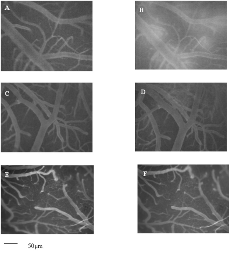
Fig 3. Computer-assisted images of hamster pial microvascular networks.
Computer-assisted image of a pial microvascular network under baseline conditions (A) and at the end of reperfusion (B) in a hamster subjected to BCCAO and reperfusion. The increase in microvascular leakage is outlined by the marked change in the color of interstitium (from black to white). Computer-assisted images of a pial microvascular network under baseline conditions (C, E) and at the end of reperfusion in a Vaccinium myrtillus supplemented diet-fed hamster for four (D) and six months (F), where the leakage of fluorescent-dextran was significantly reduced. Scale bar = ___ 50μm

