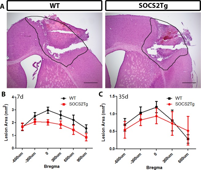
Fig 2. SOCS2Tg mice had a smaller lesion size than WT mice 7d post moderately-severe TBI.
The lesion area of injured SOCS2Tg and WT mice was assessed by haematoxylin and eosin staining of coronal tissue sections. Representative images, indicating the traced lesion area, from injured WT and SOCS2Tg mice are shown (A). At 7d SOCS2Tg mice had a smaller lesion, with a significant effect of genotype on lesion area between genotypes (F(1,73) = 12; P = 0.0008; Two-way ANOVA) but no significant differences were found in post hoc analysis (B). By 35d the lesion area was smaller than at 7d and there was no significant difference between genotypes. Results show mean area ± SEM; n = 5–8 mice/group (B, C). Scale bars in A = 500μm.

