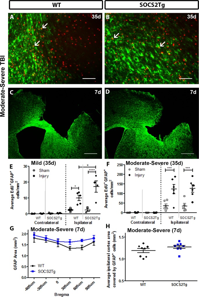
Fig 6. SOCS2 overexpression increased numbers of proliferative astrocytes in injured cortex compared to WT after mild but not moderately severe TBI.
The perilesional cortex of SOCS2Tg and WT mice was analysed at 7d and 35d post TBI or after sham surgery for density of EdU+GFAP+ cells. Representative images of WT (A, C) and SOCS2Tg (B, D) cortex are shown. Scale bar A,B = 100 μm; C,D = 500 μm. Examples of co-labelled cells in injured cortex after moderately severe TBI are indicated by arrows in panels A and B and low power images used for thresholding cortical area covered by GFAP at 7d post-TBI are in panels C and D. In the ipsilateral cortex of SOCS2Tg and WT injured animals there was an overall injury-induced increase in EdU+GFAP+ cell density at 35d post mild (E) and moderately severe TBI (F), which was further enhanced in SOCS2Tg mice after mild TBI (E). At 7d after moderately severe TBI, the area covered by GFAP expression was measured by thresholding using ImageJ. There was an overall increase in GFAP area in SOCS2Tg mice when measured across Bregma levels (G) which was not significant when sections from each animal were combined and averaged (H). Results show mean ± SEM; n = 5–8 mice/group (E-H); *P<0.05, **** P<0.0001 (ANOVA with Bonferroni post hoc test).

