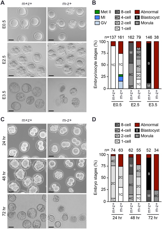
Fig 7. Embryos lacking maternal Setdb1 exhibit progressive developmental delays and fail to develop to blastocysts.
(A, B) Superovulated control and Setdb1 KO females were mated with WT males, and embryos (as well as unfertilized GV, MI, and Met II oocytes) were collected at E0.5, E2.5, and E3.5, respectively, and their developmental stages determined by morphologies. Shown are representative images (A) and percentages of embryos, as well as unfertilized oocytes, at different stages (B). Abnormal embryos/oocytes included those exhibiting abnormal morphologies and undergoing degeneration. The total numbers of embryos/oocytes examined for each genotype at each time point are indicated. (C, D). Embryo culture in vitro. Superovulated control and Setdb1 KO females were mated with WT males, and morphologically normal Setdb1m+z+ and Setdb1m-z+ zygotes were isolated at E0.5. The embryos were cultured for 24, 48, and 72 hours in vitro, and their developmental stages determined by morphologies. Shown are representative images (C) and percentages of embryos at different stages (D). The total numbers of embryos examined for each genotype at each time point are indicated.

