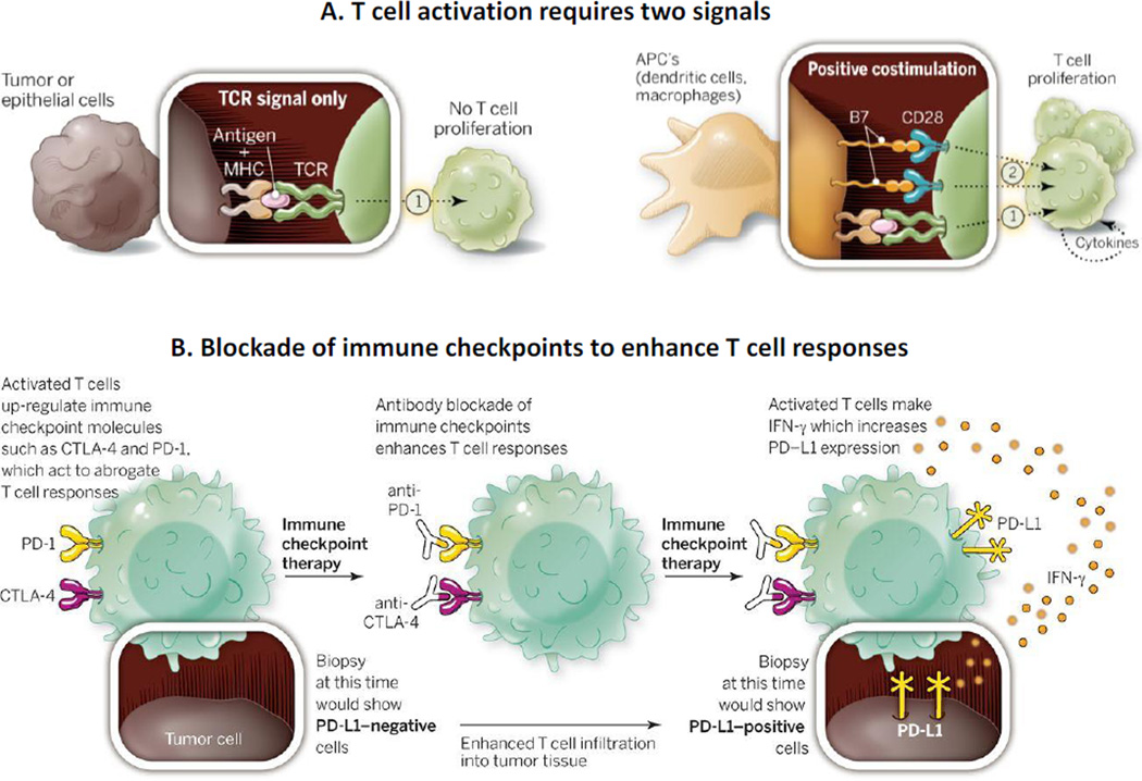
Figure 4. Immune checkpoint therapy: Pharmacological basis.
A. T cell activation requires two signals. T cells recognize tumor antigens presented by the major histocompatibility complex (MHC) on the surface of cells through their T-cell receptor (TCR). This first signal (1) is insufficient to turn on a T-cell response, and a second signal (2) delivered by the B7 costimulatory molecules on antigen presenting cells (APC) is required for activation. B. Blockade of immune checkpoints to enhance T cell responses. After activation, T cells express immune checkpoints such as CTLA-4 and PD-1. They further secrete IFN-γ, which leads to expression of PD-L1 on tumor cells and inflammatory cells and causes inhibition of T cells upon interaction with PD-1. Blocking of immune checkpoints with antibodies prevents T cell inactivation and enhances T cell responses. The expression of PD-L1 on tumor cells may be absent in early biopsies obtained prior to immune checkpoint therapy but would be detectable after the therapy. Figure and legend are reprinted with permission from [221]. The legend was adjusted for the current discussion.

