
Figure 7.
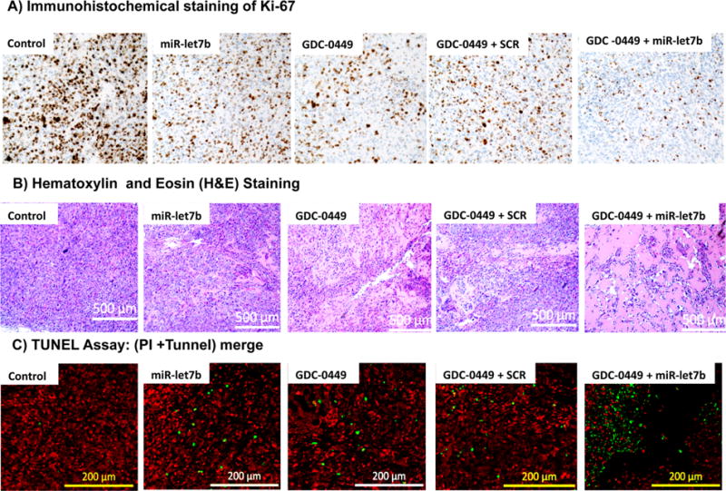
Analysis of tumor samples for Ki-67 staining, hematoxylin and eosin (H&E) staining, and (C) TUNEL assay for apoptosis. (A) Immunohistochemical staining of Ki-67, (B) H&E staining of peripheral tumor regions, and (C) apoptosis in tumor cells as indicated by the green fluorescence of TUNEL, while red spots mark the cells with propidium iodide (PI) staining.