
Figure 7.
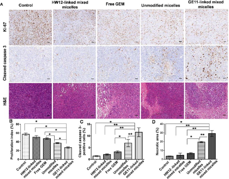
Immunohistochemical analysis of tumor samples for Ki-67 (cell proliferation marker), cleaved caspase 3 (apoptosis marker), and H&E staining (A). Saline, free GEM, HW12-linked mixed micelles, unmodified micelles, and GE11-linked mixed micelle-treated tumor samples were cryosectioned, fixed, and immunostained for Ki-67, cleaved caspase 3, and H&E (A). Statistical analysis was performed for Ki-67 (B), *p < 0.001; cleaved caspase 3 (C), *p < 0.05; **p < 0.01 and H&E (D), *p < 0.01; **p < 0.001. Scale bar, 2 mm.