
Figure 6.
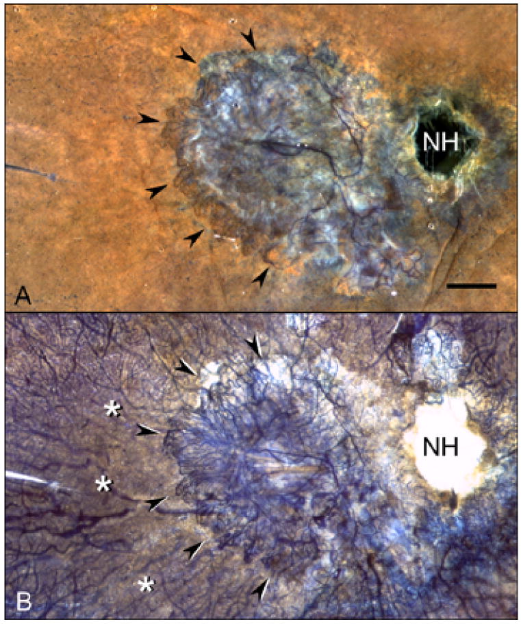
APase choroid from an 81 year-old Caucasian female with wet AMD (Case #10). A submacular seafan-like CNV formation is shown (arrowheads) using epi-illumination (A) and transillumination (B). Areas of choriocapillaris dropout are located in advance of the CNV (asterisks). Percent RPE and vascular area measurements made in 2 mm intervals from the CNV (C) show that capillary dropout is present well beyond the extent of neovascularization. (NH = nerve head, scale bar = 2 mm)