
Figure 6.
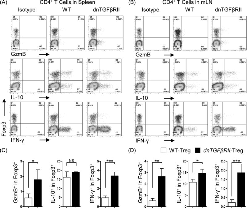
Comparative analysis of cytokine secreting capacity between dnTGFβRII;Foxp3GFP mice Tregs and WT;Foxp3GFP mice derived Tregs.
Total mononuclear cells from spleen (A) and mesenteric lymph nodes (B) of dnTGFβRII;Foxp3GFP and WT;Foxp3GFP mice were stimulated with PMA and ionomycin for 3 hours in the presence of Golgi stop reagent. Secreted IFN-γ, IL-10 and Granzyme B (GzmB) of Tregs were assessed by flow cytometry. Graphs present 10 week-old mice, 4 mice per group. (C) and (D) present the statistics analysis of the data indicated in (A) and (B), respectively. Graphs present mean ± SD. Data is representative of two independent experiments with similar results. *P <0.05, **P <0.01 and ***P <0.001 as determined by Student Test.