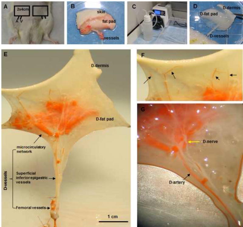
Fig. 1. Perfusion decellularization and flap matrix angiography.

(A, B) Groin skin/adipose tissue flaps with vascular pedicles (2 cm × 4 cm) were harvested from Fischer 344 rats. (C, D) Transparent DSAF matrix was achieved using a bump perfusion system combined with agitation decellularization. (“D-” indicates “decellularized” in this and other figures.) (E) Microfil-117 angiography showed that DSAF retained femoral vessels, superficial inferior epigastric vessels, and microcirculatory vessels. (F) Artery perforators penetrated into the dermis (black arrows). (G) An acellular sensory nerve (yellow arrow) and artery (black arrow) are shown.
