
Fig. 2. Characterization of DSAF.
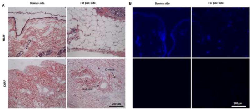
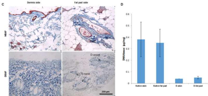
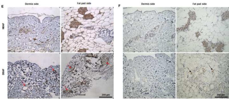
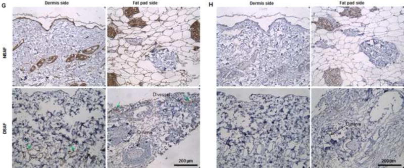
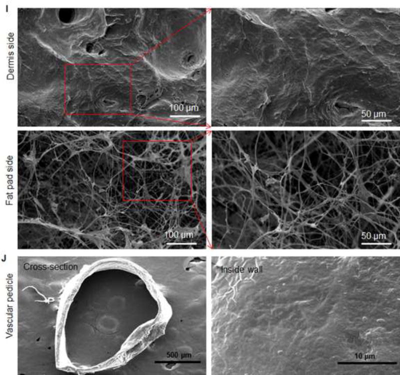
(A) H&E staining showed that blood vessels and nerve structures were well maintained in DSAF. Cell nuclei were present in NSAF but absent in DSAF. (B) DAPI staining revealed the absence of nuclei DNA in DSAF. (C) Masson trichrome staining showed that collagen was a major component of DSAF. (D) The DNA content in the decellularized skin and fat pad was significantly lower than that in the native skin and fat pad (P<0.05). (E) IHC analysis indicated that laminin was distributed in vessels, nerves, and nanofibrous structures in DSAF (red arrows). (F) bFGF was present in the glandular and nanofibrous structures of DSAF (black arrows). (G) VEGF was present in the vessels and nerve structures of DSAF (green arrows). (H) The absence of MHC-I indicated the removal of alloantigenicity from DSAF. (I) SEM images confirmed that cells were absent in DSAF, leaving 3D porous structures in the fat pad side. Nanofibrous structures of ECM were well maintained in DSAF. (J) SEM images showed that the decellularized femoral artery remained elastic and patent.