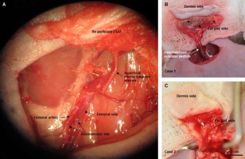
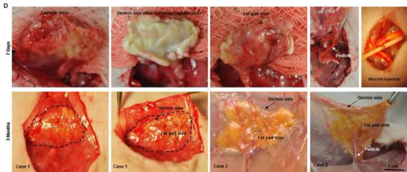
Fig. 4. Implantation and explantation of the engineered DSAF-cells construct in group A.


(A) The vascular pedicle was re-integrated into the host using conventional microsurgical anastomosis techniques. (B, C) Blood immediately perfused the engineered flap construct. (D) At 7 days (top row), the implant was encapsulated in swollen tissue (first image). Removing the swollen tissue capsule from the fat pad resulted in bleeding (second and third images). Mircofil-117 went through the vascular pedicle (fourth image) when it was injected through the contralateral femoral artery at day 7 (fifth image). At 3 months (bottom row), the implant was remodeled. Adipose tissue formed on both the dermis side and fat pad side. Numerous visible blood vessels penetrated into the newly formed soft tissue, indicating that it was highly vascularized.
