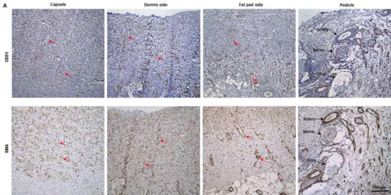
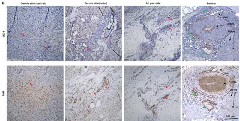
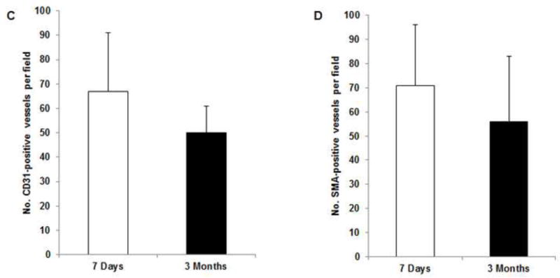
Fig. 6. IHC analysis of explants in group A.


(A) Abundant CD31- and SMA-positive blood vessels were distributed in the capsule, dermis, and fat pad area of the explant (red arrows) at day 7. (B) The adipose tissue was highly vascularized, showing numerous CD31- and SMA-positive vessels (red arrows) throughout the explant at 3 months; lots of red blood cells were present in the functional vessels. Although the main pedicle artery showed myointimal hyperplasia, lots of vessels grew in the adventitia area (green arrows). (C&D) The numbers of CD31-positive vessels and SMA-positive vessels at 7 days and 3 months were both large and did not differ significantly.
