Figure 1.
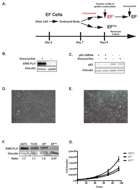
EWS-FLI1 expression in embryoid bodies. (A) Schematic diagram illustrating the differentiation protocol of the human embryonic stem cells. The culture protocol consists of the three steps: 1) embryoid body formation under non-adherent conditions; 2) transfer of embryoid bodies to gelatin-coated plates; 3) monolayer culture of the cells that outgrow from the embryoid bodies. Doxycycline is added to the embryoid bodies after seven days of culture. (B) Western blot analysis showing the inducible expression of EWS-FLI1 in the embryoid bodies in the presence, but not absence, of doxycycline. (C) Western blot analysis showing the constitutive knockdown of p53 by an shRNA in embryoid bodies. Knockdown efficiency is not affected by doxycycline. (D) EFFib cells exhibit a morphology similar to fibroblast cells. (E) The EF+ cells exhibit a distinct morphology and are more rounded and less elongated than the EFFib cells. (F) Western blot analysis of EWS-FLI1 expression in Ewing sarcoma cell lines (A673 and TC32), EF+ cells and EFFib cells. The relative expression level of EWS-FLI1 for each cell line compared to EF+ is shown below the blot. (G) The growth rate of the EF+, EFFib and A673 cells was measured using trypan blue exclusion and cell counting. The data were fit to an exponential curve using GraphPad Prism. Error bars indicate the standard deviation of three replicates.

