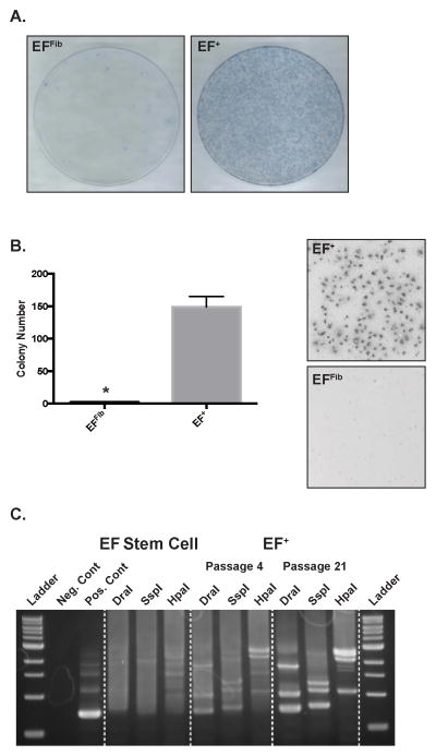
Figure 4.
The EF+ cells exhibit properties of transformation. (A) The EFFib and EF+ cells were cultured for fourteen days without passaging and then colonies were stained with methylene blue. (B) Soft-agar assay for anchorage-independent growth of EFFib and EF+ cells. Error bars indicate the standard error of the mean of three experiments (*, p<0.05). (C) Lentiviral integration analysis. Genomic DNA was isolated from the parental stem cells and EF+ cells at passage 4 and 21. Integration sites were then analyzed in three digestion libraries using a genome walking approach.

