
Figure 2.
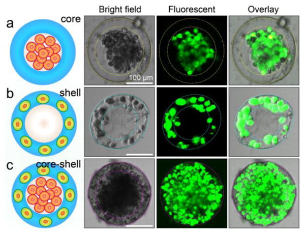
Spatial assembly of different cells in the 3D core-shell scaffold. a) HepG2 cells confined in the core by the hydrogel shell. b) NIH-3T3 fibroblasts immobilized by the cross-linked alginate network in the shell. c) Simultaneous assembly of hepatocytes in the core and fibroblasts in the shell, forming an artificial liver in a drop. Cell viability is characterized by Calcein AM/EthD-1 staining kit. The scale bars are 100 μm.