
Figure 3.
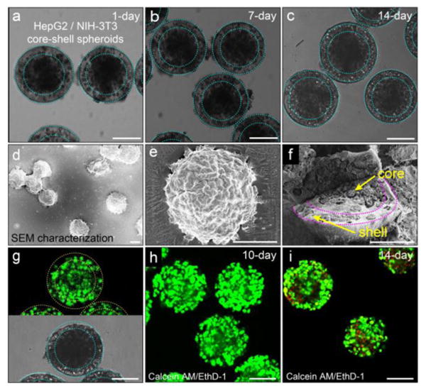
Co-culture of hepatocytes and fibroblasts in the core-shell spheroids. Morphology of the heterocellular spheroids and viability of the cells in the hydrogel scaffold. a-c) Co-culture of hepatocytes in the core and fibroblasts in the shell for 1 day, 7 days and 14 days, respectively. The heterocellular spheroids maintain their spherical morphology over time. d) SEM image and e) magnified image show individual spheroids that maintain their structural integrity after freeze-drying. f) Spatially confined cell ensembles in the core-shell structure. g) High viability of cells encapsulated in the spheroids after being frozen at -80°C for two weeks and then thawed at 37°C. Cells are predominantly alive (green) and there is complete lack of dead cells (red). h) Co-culture of cells encapsulated in the spheroids for 10 days shows high cell viability. i) The viability of cells cultured for 14 days decreases slightly, as evidenced by the appearance of dead cells (red). Cell viability is characterized by Calcein AM/EthD-1 staining kit. The scale bar is 100 μm in all images.