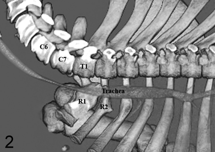
Fig. 2.
Three-dimensional reconstruction image of CT in Case 1, showing tracheal collapse and stenosis at the sixth cervical to first thoracic vertebrae (left lateral view after removal of left scapular, forelimb and ribs). Fractured marks are also recognized in the right first (R1) and second (R2) ribs. C6: sixth cervical vertebra, C7: seventh cervical vertebra, T1: first thoracic vertebra.

