Figure 4.
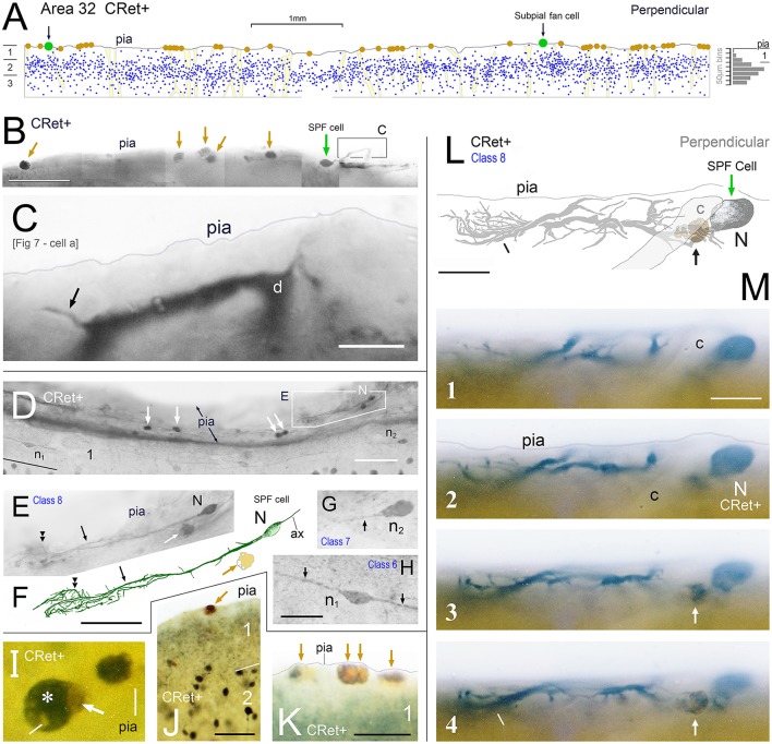
(A) Drawing of a perpendicular coronal strip through area 32 showing the location of CRet+ neurons (blue dots) in layers 1, 2, and upper 3. Subpial thorn (SPF) cells (green dots). Subpial somatic profiles containing dark yellow lipofuscin (brown dots). Profiles of penetrating blood vessels (yellow). A summary depth-frequency distribution of CRet+ cells is given on the right. Scale bar: 1 mm. (B) Photographic montage of the pial surface of a coronal section through area 46v. A SPF cell is indicated with part of its dendritic arbor highlighted. Note discrete dark cellular profiles (brown arrows). Scale bar: 100 μm. (C) Part of the main immunolabeled dendrite (d) of the SPF cell in (B). A dendritic thorn is indicated (arrow). Scale bar: 10 μm. [The morphology of this SPF cell is shown in Figure 7–cell a]. (D) Oblique view of the pial surface in area 24b showing a CRet+ SPF cell (N) and numerous dark somatic profiles containing cellular debris distributed under the pia (white arrows). Note also two CRet+ cells in mid-layer 1. Scale bar: 100 μm. (E) Class 8. Enlargement of the SPF cell in (D). Scale bar: 100 μm. (F) Drawing of the dendritic arbor of the SPF in (D,E). Axon, ax. (Subpial lipofuscin-rich profile — brown). Scale bar: 50 μm. (G,H) Magnified views of CRet+ cells n1 and n2 in (D). Cell n1 is a deep horizontal bipolar cell (Class 6) and n2 a simple unipolar neuron (Class 7). Dendrites (arrows). Scale bar: 20 μm. (I) View of pial surface showing a vacuolated subpial CRet+ profile (gray immunolabeling — asterisk) with a large brown swelling (white arrow). Scale bar: 10 μm. (J) Area 25. Large brown subpial profile (arrow). Scale bar: 100 μm. (K) Cluster of brown subpial profiles in area 9. Scale bar: 50 μm. (L) Perpendicular view of a SPF cell (N) in area 46v. The outline of a descending capillary is indicated (c). Cellular debris containing lipofuscin (arrow). Terminal dendritic plexus (line). Scale bar: 20 μm. (M) Series of micrographs (1–4) taken at various focal planes through tissue section containing the CRet+ SPF cell indicated in (L). Capillary (c); deposit of cellular debris associated with labeled dendrite (arrow); and terminal dendritic plexus (line). Scale bar: 20 μm.

