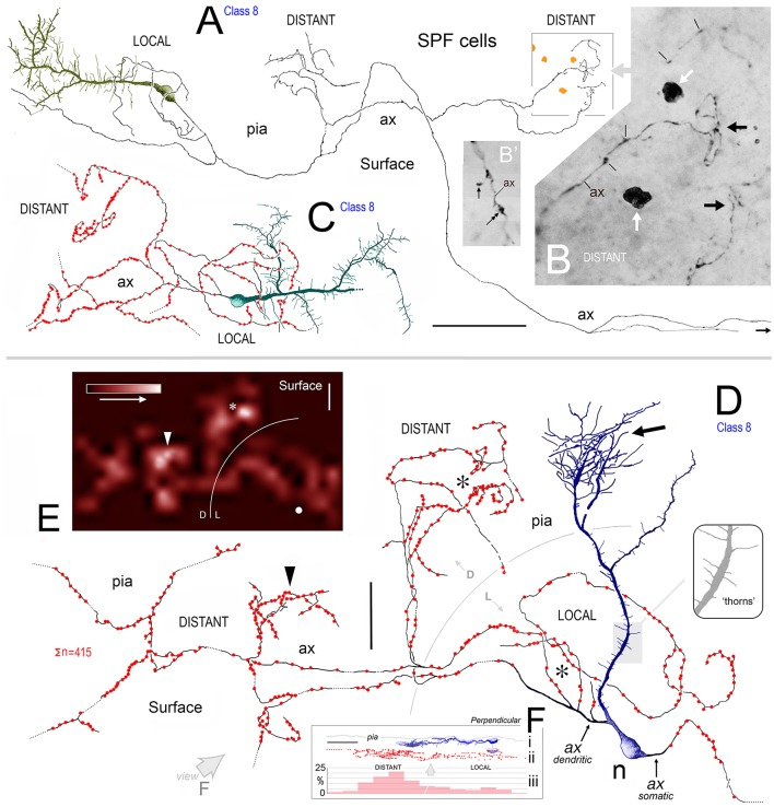
Figure 8.
(A) View of pial surface in area 32. A class 8 SPF cell gives rise to a tangential axonal plexus that ramifies both proximally (LOCAL) and distally (DISTANT) to the soma. (B) Boxed region in (A) showing numerous varicosities (arrows) along two distal segments of the axon (ax). Dark irregular profiles of cellular debris (white arrows). (B′) Axonal boutons — terminaux (arrow) and en passant (double headed arrow). (C) Surface view of a class 8 SPF cell in area 9. Individual axonal swellings along the proximal and distal sections of the axon are indicated (red dots). Note fan-like dendritic trees of both SPF cells. Scale bar for (A,C): 100 μm. (D) Tangential view of a class 8 SPF cell (n) in area 24b with a fine varicose axonal process (ax) emerging from one somatic pole (ax somatic). The other pole gives rise to a proximally bifurcating thick primary dendrite with numerous dendritic “thorns” (shaded region and inset) and terminates in a fan-like plexus of fine processes (arrow). A proximal dendritic branch bifurcates into several fine varicose axonal fibers (ax dendritic) that ramify extensively — both locally and at a distance from the soma. Boundary (150 μm radius from soma) between LOCAL (L) and DISTANT (D) zones is indicated. The morphologies of these processes are similar to those seen in (A,C). Scale bar: 50 μm. (E) Density distribution of axonal varicosities from neuron in (D). (Lattice resolution 20 × 20 μm squares). White arrow head indicates cluster of varicosities indicated in (D). Soma (white dot). Scale bar: 50 μm. (F) Perpendicular view of thorn cell viewed along axis indicated (open arrow in D). (i) dendritic arbor, (ii) distribution of axonal varicosities relative to somatic profile, and (iii) frequency of axonal varicosities within 50 μm wide sampling bins — note distribution in distant and local zones. Scale bar: 100 μm.

