Figure 6.

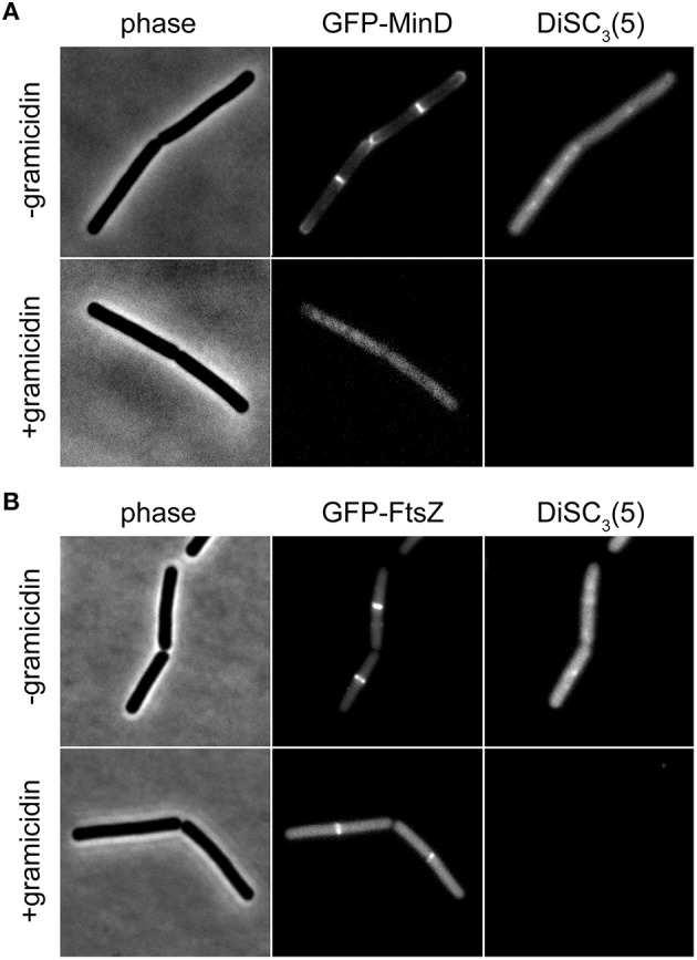
Compatibility of DiSC3(5) and GFP in microscopic single-cell experiments. (A) Phase contrast image (left panel) of cells expressing GFP-MinD (middle panel) and stained with DiSC3(5) (right panel) in the absence and presence of gramicidin (5 μM). Note the strong decrease in DiSC3(5) fluorescence and delocalization of MinD upon depolarization. (B) Phase contrast image (left panel) of cells expressing GFP-FtsZ (middle panel) and stained with DiSC3(5) (right panel) in the absence and presence of gramicidin (5 μM). Note the reduced septal signal of FtsZ upon depolarization. Strains used: B. subtilis KS64 (GFP-MinD), and B. subtilis 2020 (GFP-FtsZ).
