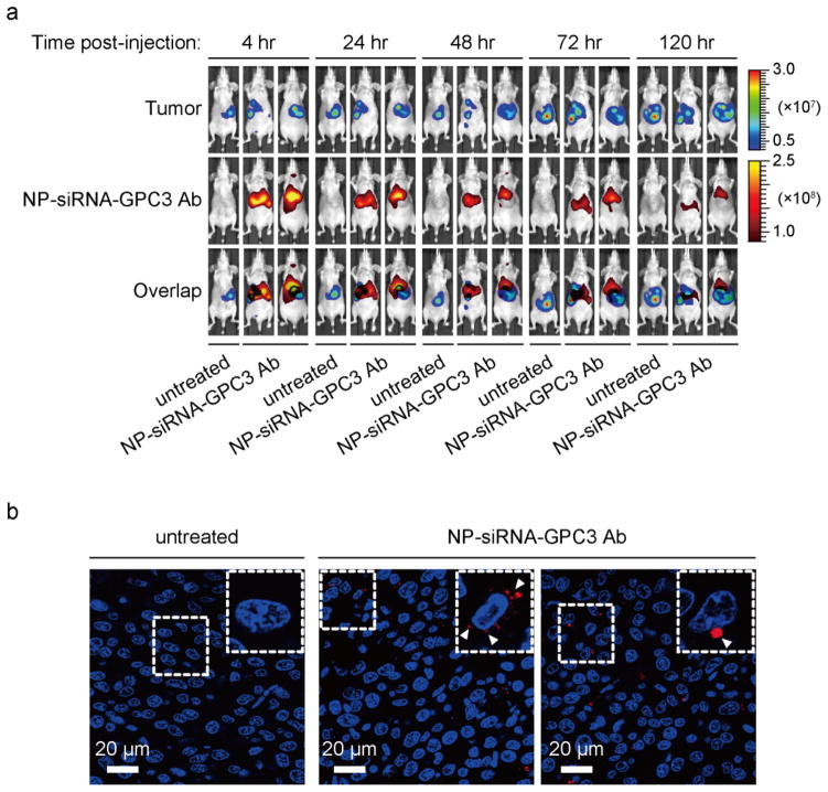
Figure 6.
Evaluation of tumor uptake of NP-siRNA-GPC3 Ab in vivo. (a) Xenogen IVIS images showing co-localization of RH7777-Luc-GPC3 tumor and Dy677-labeled siRNA loaded NP at different time points post-injection from one untreated and two treated mice. (b) Confocal fluorescence microscopy images showing accumulation of NP-siRNA-GPC3 Ab in tumor sections at 120 hr after injection from the same animals presented in panel (a).

