Figure 5.
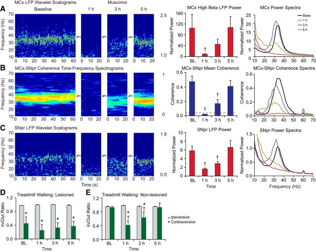
Effect of muscimol infusion into the VM thalamus on the LFP oscillations in the MCx and SNpr in control and dopamine cell-lesioned rats during treadmill walking. A–C, Wavelet-based scalograms of LFP spectral power (left) in the MCx (A) and SNpr (C) and the MCx–SNpr FFT-based coherence (B). Recordings were made during baseline (BL) treadmill walking and after 1–5 h after infusion of muscimol into the VM in the lesioned hemisphere (n = 6, 40 ng/0.5 μl). Bar graphs (middle) show MCx and SNpr LFP total normalized power and their coherence (mean ± SEM) before and after 1–5 h of muscimol administration into the VM. The plots and scalograms indicate that infusion of muscimol into the VM significantly reduces MCx and SNpr 30–36 Hz range LFP power (p < 0.05) and MCx–SNpr coherence (p < 0.001) in the lesioned hemisphere at 1 h. LFP power and MCx–SNpr coherence gradually returns to baseline levels over 5 h after treatment. Plots in the right panel represent averaged power spectra and MCx–SNpr coherence before and after muscimol infusion. D, E, Bar graphs representing step count ratios for the inner to outer hind limbs during ipsiversive (gray bars) and contraversive (green bars) walking in lesioned (D) and control (E) rats before and after infusion of muscimol. Infusion of muscimol into the VM does not notably alter impaired gait in lesioned rats. Muscimol injected into the left VM in the control rats (n = 4) dramatically reduces step ratios in contraversive walking at 1 and 3 h after injection (p < 0.003). *Significant difference between the ratio of steps in contraversive relative to ipsiversive walking. †Significant difference from baseline.

