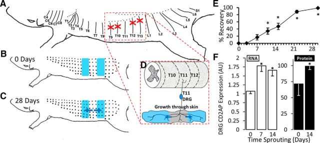
Figure 1.
CD2AP is upregulated during sensory arbor expansion in the spared dermatome model. A, T9, T10, T12, and T13 DCNs were transected and the proximal cut-end ligated to prevent regeneration (red crosses). B, Denervated zones (shaded blue in B–D) were mapped by pinching the skin with fine forceps to drive the CTMR. B, C, Black dots indicate responsive sites. C, Arbor expansion proceeds from the spared fibers into the denervated zones. Blue arrows indicate direction of arbor expansion. D, Dermatome-focused (segmental) schematic of arbor expansion of cutaneous projections from the T11 DRG. Growth is in the direction of the blue arrows and proceeds into the denervated zones (shaded blue). E, Time course of arbor expansion determined using the CTMR. Size of the denervated zones is recorded at time 0 (baseline) until complete reinnervation. Reinnervation of the entire denervated area takes ∼28 d. *Significantly changed from day 0 (repeated-measures ANOVA, post hoc Tukey HSD). F, Expression of CD2AP mRNA (measured by qPCR) and protein (measured by Western blot) during key stages of arbor expansion. For mRNA, the control is set to a value of 1. For protein, the strongest signal was set to a value of 100. 7 d, Initiation; 14 d, maintenance. *Statistically significant changes from 0 d (naive) (ANOVA followed by post hoc t test).

