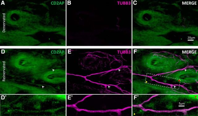
Figure 12.
CD2AP is present in growing axons in reinnervated skin. Representative maximum-intensity projection through the epidermis of denervated (A–C), and adjacent collaterally reinnervated (D–F) adult mouse dorsal skin, stained to visualize CD2AP (green) and neuron-specific tubulin (TUBB3; magenta). Skin was cleared using organic solvent to allow confocal imaging throughout the tissue in whole mount. Note the CD2AP+ axons in reinnervating skin (white arrowheads). F, Boxed area is shown in D′–F′. Colored dots indicate registration marks.

