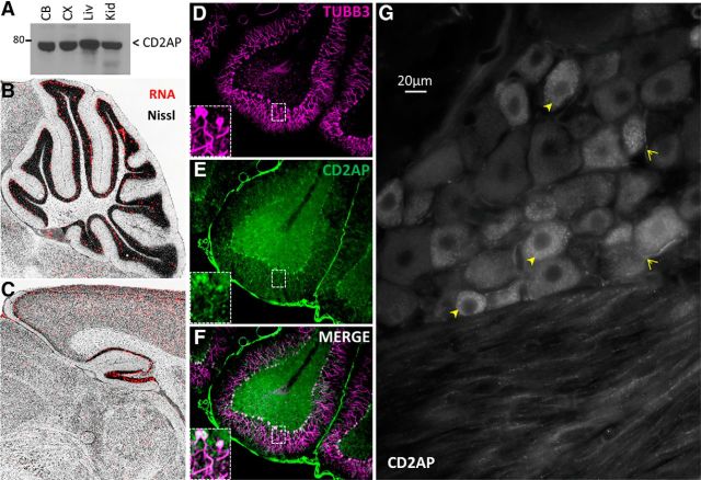
Figure 3.
CD2AP is expressed in neurons. A, Western blot assessment of CD2AP protein in tissue lysates. CB, Cerebellum; CX, cortex; Liv, liver; Kid, kidney. Allen Brain Atlas demonstrates CD2AP mRNA in cerebellum (B) and hippocampus (C). D–F, CD2AP protein expression in rat cerebellum. Note high expression levels in neurons. G, CD2AP protein in DRG. Note elevated levels of punctate staining in small diameter soma (closed arrowheads) and axons (open arrowheads).

