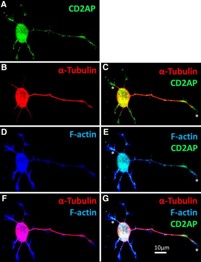
Figure 7.

CD2AP locates to the growing tips of PC12 neurites. PC12 cells were treated with NGF for 48 h, fixed, and costained to visualize the following: A, CD2AP (green); B, C, α-tubulin (red); and D, E, F-actin (using phalloidin; blue). CD2AP locates to tubulin−/F-actin+ neurite tips (F, G). *CD2AP-positive neurite tip (growth cone).
