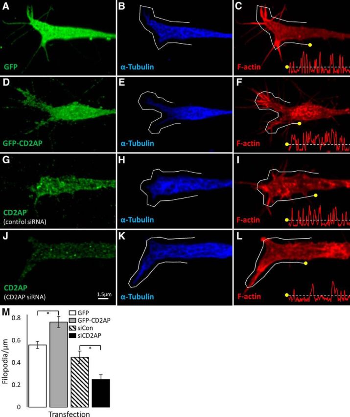
Figure 8.

CD2AP is a positive regulator of the number of filopodia on growth cones. PC12 cells were transfected with GFP (A–C), GFP-CD2AP fusion protein (D–F), control siRNA (G–I), or CD2AP siRNA (J–L) for 24 h before NGF treatment for 48 h. Cells were fixed and stained to visualize F-actin (using phalloidin; red; C, F, I, L), α-tubulin (blue; B, E, H, K), and either GFP (green; A, D) or CD2AP (green; G, J). Growth cones were imaged by confocal microscopy using 100× objective with 5× digital zoom. Images are representative deconvolved maximum-intensity projections. Line histograms were generated for lines exactly 20 μm in length applied exactly 1 μm outside of the tubulin-positive boundary (gray lines). Registration marks (yellow dots) indicate how the histogram relates to the line (x axes = distance, y axes = log of actin fluorescence intensity). M, Mean number of filopodia per condition. *Significant difference (t test). Error bars indicate SEM.
