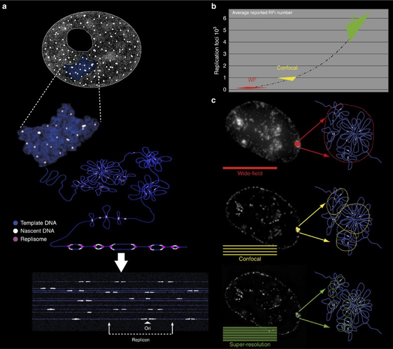
Figure 4. Replication sites dissected by super-resolution microscopy in the mammalian nucleus correspond to individual replicons.
(a) A cartoon showing how replication sites/units can be seen at different levels of chromatin compaction from the extended DNA fibres to the 3D-preserved whole-cell (nucleus) level. (b) Increase in RFi numbers driven by resolution improvements in microscopy during the past three decades42. WF: wide field; SR: super-resolution microscopy. (c) Microscopic images and corresponding cartoon interpretation of replication sites in the mammalian nucleus imaged at different levels of resolution. For summary of experimental numbers and calculations see Table 5.

