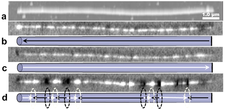
Figure 9.
Topography (a) and MFM images (b–d) of a polycrystalline core-shell nanowire (20 min annealing). (b,c) at remanence after saturation in opposite directions parallel to the length of the nanowire. (d) Multi-domain state at 1.05 kOe external magnetic field. The schematics in (b–d) show the magnetization direction (arrows) and domain wall positions (ellipses).

