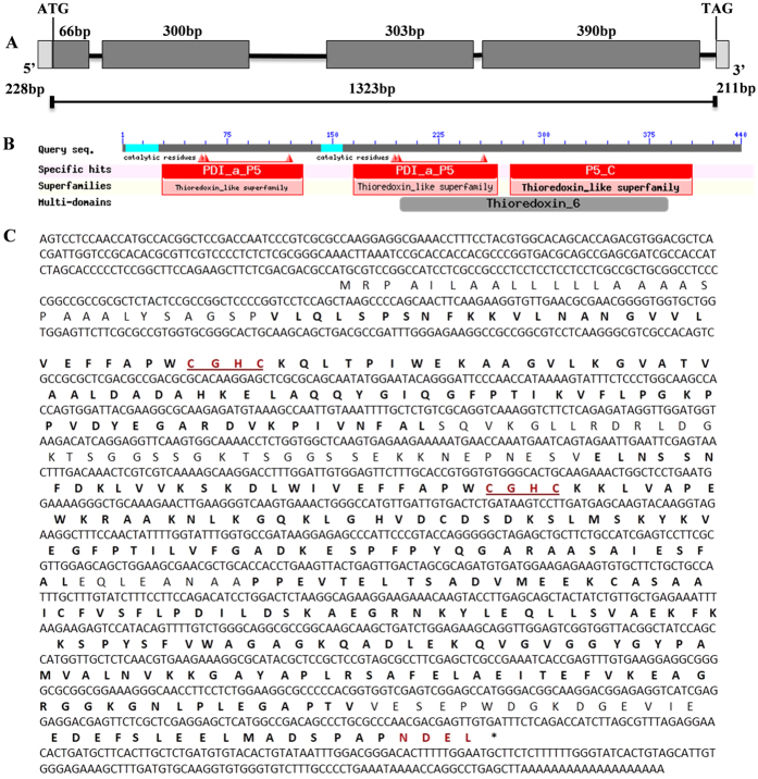
Figure 1. Structural features of the PDI-V cDNA sequence and its translation product.
(A) Structure deduced from the full-length cDNA sequence of PDI-V. The size of each domain, signal peptide and 3′ and 5′- UTR sizes and position of the ATG and TAG stop codons are shown. (B) Graphical representation of domain organization as provided by the NCBI conserved domain database. (C) Deduced amino acid sequence of PDI-V. Thioredoxin catalytic motifs and ER retention signals are shown in red.

