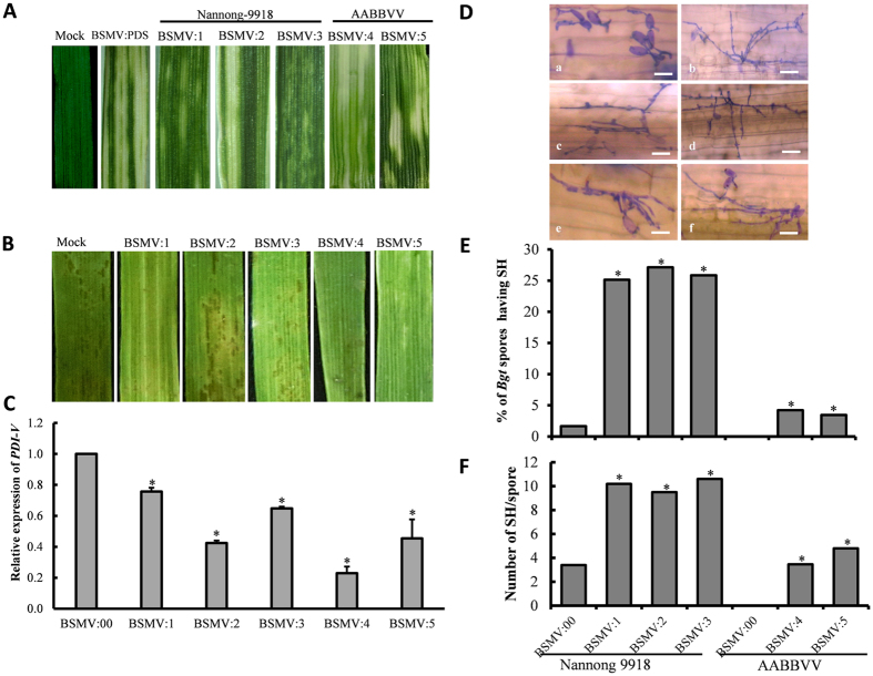
Figure 6. Functional characterization of PDI-V in Bgt-infected Nannong 9918 and H. villosa-T. turgidum amphiploid (AABBVV) by BSMV-mediated VIGS.
(A) Typical photo bleaching of BSMV:PDS-infected was first observed on the third leaf at 9 dpi. Photographs show the fourth leaves at 15 dpi. (B) Disease responses at 7 dpi of Bgt inoculation on the fourth leaves of BSMV:PDI-V-silenced plants. (C) Assessment of silencing efficiency of BSMV:PDI-V by qRT-PCR assay. The fifth leaves of PDI-V-silenced plants were sampled; BSMV:00 infected plant were used as the control. Data were normalized using the Tubulin gene as an internal control. Each point represents the mean of three replicates. Bars indicate SD, *p < 0.05. (D) Microscopic observation of fungal growth in BSMV:00 (a) and BSMV:PDI-V infected plants (b–d from Nannong 9918; e,f from the AABBVV amphiploid). Scale bar = 30 μm (E) Comparison of Bgt spores producing SH between BSMV:00- and BSMV:PDI- infected plants (BSMV:1 to BSMV:3, Nannong 9918; BSMV:4 and BSMV:5, AABBVV). (F) Numbers of individual fungal spores producing secondary hyphae were counted from whole detached leaves of each genotype and compared with BSMV:00. *P < 0.05 according to Student’s t-test.

