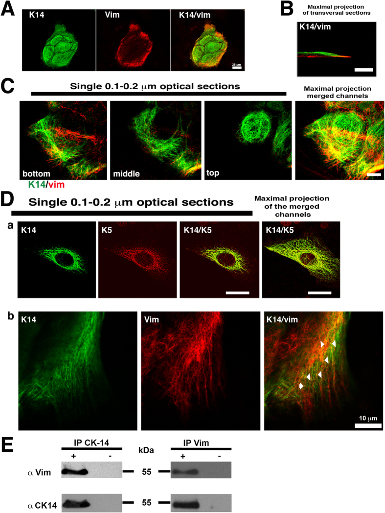
Figure 1. Vimentin and keratin stained in cultured human epidermal keratinocytes.
(A) Immunofluorescence staining of a 4 day-old human keratinocyte colony for KRT14 keratin (green) and Vim (red). Note that only peripheral cells expressed Vim in addition to keratins. (B) Confocal microscopy image of a keratinocyte immunostained for KRT14 (green) and Vim (red). (C) Serial single optical sections by confocal microscopy showing the immunofluorescence staining for KRT14 and Vim at different focal planes. Note that while keratin IFs form a basket-like structure encasing the nucleus, Vim is present only in basal cells at the bottom. Scale bar = 10 μm. (D) Single optical section by confocal microscopy showing the immunofluorescence staining for KRT5/KRT6, or KRT14 and Vim; in (a) Note the complete co-localization of KRT14 with KRT5, and in (b) partial co-localization of KRT14 and Vim. (E) Representative immunoblot analysis showing the co-immunoprecipitation of Vim and KRT14, (+) in presence or (−) absence of the primary antibody.

