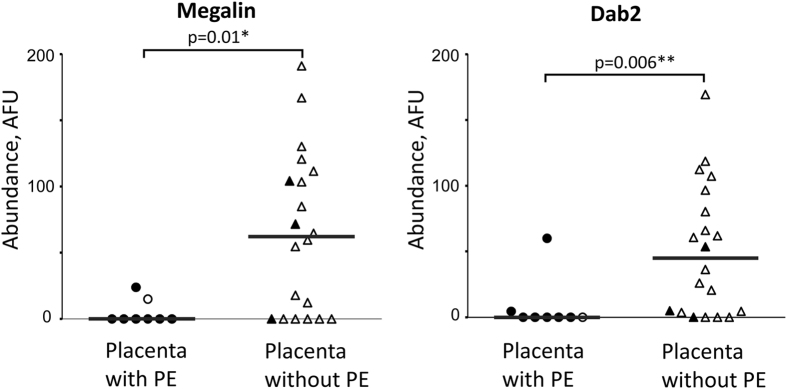
Figure 1. Megalin and Dab2 abundance is reduced in brush border area of syncytiotrophoblast in placentas with malarial infection at delivery.
Abundance of megalin and Dab2 was assessed in brush border of placentas with PE (n = 8) and without PE (n = 20) using immunofluorescence assay as described in Methods. Medians of arbitrary fluorescence units (AFU) are reported (gray bars). Filled symbols represent samples with extracellular hemozoin in fibrin indicative of previous (resolved) placental infections, clear symbols represent samples without hemozoin. Protein abundance between 2 groups was compared using Mann-Whitney test. PE – parasitized erythrocytes in the placenta at the time of delivery.

