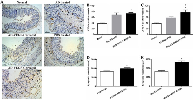
Figure 3. Immunohistochemical results for lymphatic remodeling in acute colitis (A). Comparison of lymphatic vessel density (LVD) between AD-VEGF-C treated and control mice (B) and between AD-VEGF-C156S-treated and control mice (C). AD-VEGF-C did not induce an increase in LVD (P=0.50), whereas VEGF-C156S induced a 1.3-fold increase in LVD compared to PBS-treated mice. Comparisons of lymphatic vessel size between AD-VEGF-C-treated and DSS-treated mice are shown in panel D and between AD-VEGF-C156S-treated and PBS-treated mice are shown in panel E. Both AD-VEGF-C and VEGF-C156S-treated mice had significantly greater lymphatic vessel size compared to control mice (both P<0.001). Data are reported as means±SD (n=5/group). *P<0.05 compared to DSS-treated group. †P<0.05 compared to water-treated group. Statistical analysis was performed by ANOVA and Bonferroni's post-hoc test for lymphatic vessel density and two-sample t-test for lymphatic vessel size. AD: adenovirus; PBS: phosphate-buffered saline; AD-VEGF-C: adenovirus vascular endothelial growth factor-C; DSS: dextran sodium sulfate.