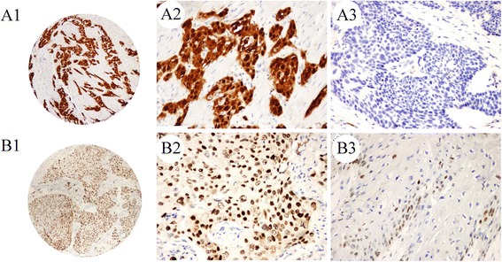
Fig. 1.

Immunohistochemical staining of p16 and p53 in Kazakh ESCC tissues. High p16 and p53 expression levels in ESCC (A1, p16; B1, p53; original magnification 40×). High power view (original magnification 200×) shows positive staining for p16 and p53 in the nucleus/cytoplasm and nucleus staining of cancer cells, respectively (A2, p16; B2, p53) and p16- and p53-negative expression (A3, p16; B3, p53; original magnification 200×)
