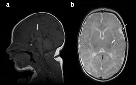
Fig. 1.

Brain MRI at age 18 days. Sagittal T1WI view showing thinning of the corpus callosum (arrow), with anterior predominance, and some degree of frontal lobe volume reduction (a). Axial T2WI view showing asymmetric enlargement of temporal subarachnoidal spaces (arrow) and mildly prominent sulci over the cerebral convexity, suggesting incipient atrophy. Myelination is delayed with minimal signals observed at the posterior limb of the internal capsule (arrowhead) and corpus callosum (b)
